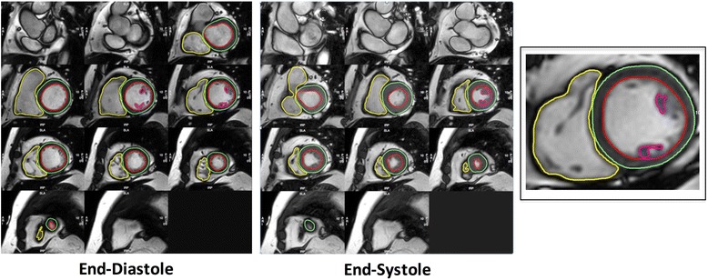
Fig. 1.

Contouring of Left and Right Ventricles in Diastole and Systole. Endo- and epicardial borders of the left ventricle were contoured in end-diastole and end-systole. In the left ventricle, papillary muscles were included in myocardial mass assessment and excluded from volumes estimation. Left ventricular mass was corroborated in end-diastole and end-systole. In the right ventricle, trabeculations and papillary muscles were included as part of the ventricular cavity so as to improve reproducibility
