Fig. 1.

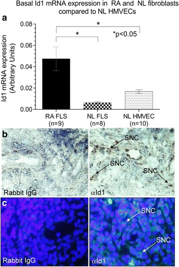
Id1 is expressed in ECs and ST fibroblasts. a mRNA was isolated from HMVECs and fibroblasts were isolated from NL and RA ST. mRNA was reverse transcribed into cDNA and underwent PCR for 40 cycles. RA fibroblasts showed significantly elevated Id1 expression compared to NL ST fibroblasts and HMVECs. b Id1 is expressed in RA STs. IHC was performed on RA, OA, and NL ST cryosections. Tissues were blocked and then incubated using a mouse anti-human Id1 (Abcam) primary antibody. After washing, tissues were incubated with a biotinylated anti-mouse secondary antibody (Vector Laboratories). Tissues were washed and subsequently developed with the Vectastain ABC kit (Vector Laboratories). Id1 is found on synovial cells (SNC) in the RA ST. c For immunofluorescence staining, RA, osteoarthritis (OA), and normal (NL) ST sections were fixed in cold acetone for 30 min. The STs were blocked with 5 % donkey serum and 20 % fetal bovine serum (FBS) in PBS at 37 °C for 1 h, and then incubated with rabbit anti-human Id1 antibody (Abcam, 10 μg/ml) or purified nonspecific rabbit IgG for 1 h at 37 °C in blocking buffer. The synovial tissues samples were washed with PBS, and a 1:200 dilution in blocking buffer of fluorescent-conjugated donkey anti-rabbit antibody was added and incubated for an additional 1 h at 37 °C. As shown, we could validate Id1 staining in RA ST similar to what was found using IHC (×400). FLS fibroblast-like synoviocytes, HMVEC human dermal microvascular endothelial cell, Id1 inhibitor of DNA binding 1, IgG immunoglobulin, NL normal, RA rheumatoid arthritis, SNC synovial cell
