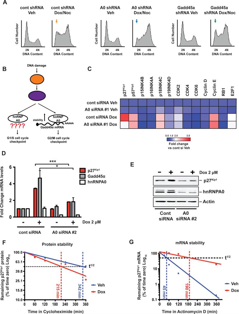
Figure 1. A focused screen implicates p27Kip1 as an hnRNPA0-dependent G1/S regulator.
(A) H1299 cells were depleted of hnRNPA0 or Gadd45α and treated with 2 μM doxorubicin for 4 hr followed by the addition of 250 ng/mL nocodazole for a further 24 hr. Cells were fixed and stained with propidium iodide and analyzed by flow cytometry. Yellow, blue and green arrows depict loss of the G1/S checkpoint in hnRNPA0 knockdown cells but not in Gadd45α knockdown cells. (B) Schematic representation of the known role of hnRNPA0 in the DNA damage response. (C) Cells were treated with doxorubicin for 16 hr, mRNA levels of the indicated targets were determined by qRT-PCR, and the data represented as fold-change vs. control siRNA vehicle-treated cells. (D) qRT-PCR analysis of H1299 cells, 16 hr post-doxorubicin treatment. Data are represented as fold change vs. control siRNA vehicle-treated cells. Error bars represent mean +/−SEM, n=3 experiments * p < 0.05, *** p < 0.001. (E) Western blots of H1299 cells 16 hr post-doxorubicin treatment. (F) H1299 cells were treated with doxorubicin for 16 hr followed by the addition of cycloheximide. Samples were taken at indicated time points and p27Kip1 protein levels measured by immuno-blotting. (G) H1299 cells were treated with doxorubicin for 16 hr followed by the addition of actinomycin D. At indicated time points p27Kip1 mRNA levels were determined by qRT-PCR.
See also Figure S1.

