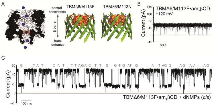
Figure 4.
Continuous nucleobase discrimination in a truncated αHL pore with a cyclodextrin adapter. (A) Left: Schematic representation of individual DNA mononucleotides (blue circles), binding inside the TBMΔ6 pore (grey, cross-section) equipped with a cyclodextrin adapter (am7βCD, orange). Right: Cartoon schematics of the TBMΔ6 β-barrel domain, showing the interaction of am7βCD (orange) with the mutants M113F (red) and M113N (purple) as determined for the untruncated pore mutants.32 (B) Representative single-channel trace of the TBMΔ6/M113F pore, in the presence of 40 μM am7βCD. The recording was made in 1 M KCl, 25 mM Tris.HCl, pH 7.5, at +120 mV. (C) Single-channel recording from the TBMΔ6/M113F•am7βCD pore showing continuous deoxyribonucleoside monophosphate (dNMP) detection (cis: 10 μM dGMP, 10 μM dAMP, 10 μM dCMP and 10 μM dTMP). Data were acquired at +120 mV. The amplified signal was low-pass filtered at 5 kHz and sampled at 25 kHz with a computer equipped with a Digidata 1440A digitizer (Molecular Devices).

