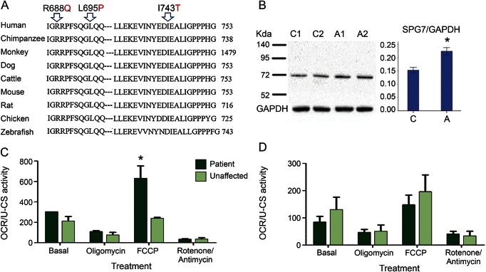
Figure 2. Increased mitochondrial sensitivity to glucose reduction in lymphoblastoid cells from patients with primary lateral sclerosis.
(A) The mutated amino acids L695 and I743 are highly conserved in vertebrate species. Mutant amino acid residues are indicated by arrows. (B) Western blot was performed using the lysates of the lymphoblastoid cells from the unaffected heterozygous (lanes 1 and 2) and the affected compound heterozygous (lanes 3 and 4) individuals in this family. GAPDH was used as a loading control. The relative ratios of band density for SPG7 over GAPDH are shown in the right panel. (C, D) Oxygen consumption rate (OCR) assays. The OCR was measured under 4 conditions: basal, complex V inhibited, uncoupler stimulated, and in the presence of complex I and III inhibitors. The lymphoblastoid cells from 3 different patients and 2 unaffected heterozygous individuals were used in normal (C) or stress (D) conditions using the standard SeaHorse protocols. In all assays, cell adhesion was facilitated by using Cell-Tak-treated assay plates. The cells were allowed to attach for 1 hour before beginning the assay. *p < 0.05.

