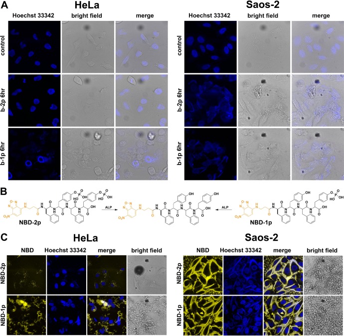
Figure 5.
(A) Nuclei staining (by Hoechst 33342) of HeLa and Saos-2 cells treated with b-2p and b-1p (500 μM) for 6 h. HeLa cells were stained for 5 min, and Saos-2 for 10 min to guarantee a clear contrast in control images. Accumulated nanofibers on cell surface trap the Hoechst 33342 and prevent this nuclei dye from entering cells. (B) Chemical structures of NBD-2p and NBD-1p (analogues of b-2p and b-1p), which turn into the same hydrogelator after dephosphorylation. (C) Confocal microscopy images of HeLa and Saos-2 cells treated with NBD-2p and NBD-1p for 12 h. Nuclei are stained by Hoechst 33342.

