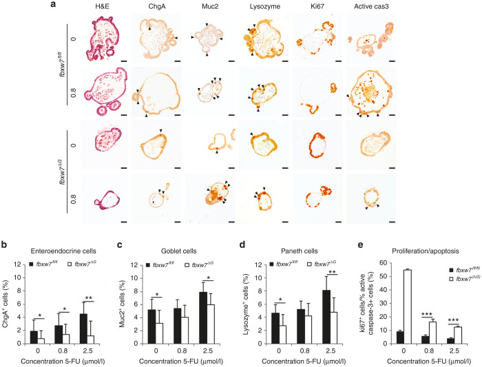
Figure 4.
Fbxw7 loss impairs 5-FU-induced differentiation and, thus, apoptosis. (a) H&E staining and immunohistochemistry (IHC) analysis of enteroendocrine cells positive for chromogranine A (chgA), goblet cells positive for mucine (muc2), Paneth cells positive for lysozyme (lys), cycling cells (Ki67), and apoptotic cells positive for active caspase 3 (active cas 3) on fbxw7fl/fl and fbxw7ΔG mini-guts following incubation with 0.8 μmol/l 5-FU. Black arrowheads show positively stained cells. Scale bars, 50 μm. (b–d) Histograms show percentage of terminally differentiated cells and ratio between cycling cells and apoptotic cells (e) following IHC analyses. The relative number of positively stained cells in 50-untreated organoids was counted for each genotype (fbxw7fl/fl versus fbxw7ΔG mini-guts) and in 30-treated organoids for each dose of 5-FU and presented as % of positively stained cells to the total number of cells (*P < 0.05, **P < 0.01, ***P < 0.001).

