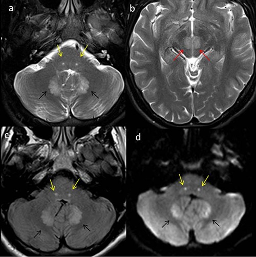
Figure 1.

Multiple axial magnetic resonance imaging of the brain in a 22-year old male on metronidazole presenting with cerebellar symptoms. Axial T2 images (a,b) reveal symmetric areas of increased signal in the dentate (black arrows), the facial (yellow arrows) and the red nuclei (red arrows), bilaterally. Axial fluid attenuated inversion recovery images (c) showing similar changes with restricted diffusion noted on the diffusion-weighted image (d).
