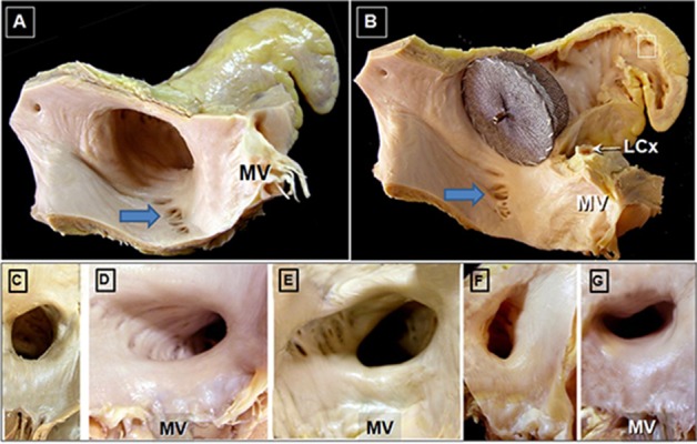
Figure 3. Morphologic Complexity and Variation in Left Atrial Appendage 3D structure Adapted with permission from Cabrera; Heart 2014 Panel A: Post-mortem specimens showing a single lobed left atrial appendage (LAA). The closely related pulmonary trunk (PT) and left superior pulmonary vein (LSPV) are also shown. Panel B: Example of a multi-lobed left atrial appendage (asteric showing distinct lobes). The aorta, as well as the pulmonary trunk and LSPV are seen in this view. Panels C-F: Examples of variant 3D morphology of left atrial appendage shape; Panel C: “Chicken Wing”, Panel D: “Windsock”, Panel E: “Cactus”, Panel F: “Cauliflower”.

