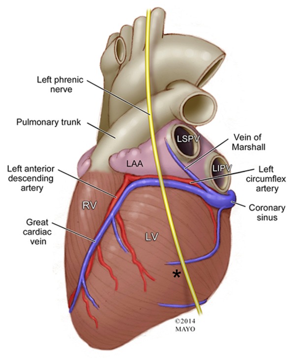
Figure 6. Endocardial and Epicardial Landmarks of the Left Atrial Appendage Ostium Top Panel: The left inset shows a gross anatomical specimen of an endocardial view of the right and left atrium transected at the annulus to show the relationship between the left atrial appendage (LAA), ligament/vein of Marshall (LOM) ridge, and coronary sinus (CS). Two probes are shown marking where the ostia of the left sided pulmonary veins are in relation to this ridge. The accompanying Illustration on the right shows a similar view of the gross anatomy after opening the LAA to provide a view the endocardial surface of the appendage. This image shows the endocardial relationship of the LOM ridge which is a marker for the overlying epicardial ligament of Marshall. This ridge separates the ostia of the left superior and left inferior pulmonary veins (LSPV and LIPV) and the ostium of the LAA. The ligament/vein of Marshall course is shown (blue-grey shadowing), as well as its connection to the CS. Bottom Panel: An illustration of an epicardial view showing the invagination of the epicardial surface which contains the vein/ ligament of Marshall is shown in the left inset. The invagination forms a boundary between the LAA and the LSPV/LIPV. The location of the epicardial invagination reflects the location of the endocardial ridge and approximation of the ostium of the LAA. The accompanying right inset shows a gross anatomical specimen depicting this view with a probe inside of the LOM.

