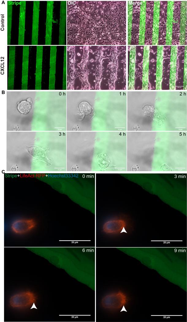
Figure 6. NPCs polarize and migrate towards CXCL12 stripes in live cell imaging experiments.
A: Mouse NPCs (1×10⁷/ml) were seeded on plastic Petri dishes (first coated with PDL) preprinted with BSA stripes (green) without CXCL12 (the control group) or with CXCL12 (CXCL12 group). After proper time, mouse NPC migrated directionally to the BSA/CXCL12 stripe. NPCs in control group showed a shape of random migration, while those in CXCL12 group displayed a shape of directional migration. B, The process of NPCs’ migration induced by CXCL12 on stripes was captured during 5 hours. C, NPCs transfected with LifeAct-RFP were polarizing towards CXCL12 stripe and forming lamellipodia. Scale bar: A, 100μm, B, 10μm, C, 20 μm.

