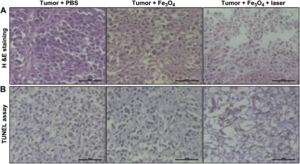
Fig. 6.
Histologic assessments of tumor tissues with and without photothermal treatments of Fe3O4/(DSPE-PEG-COOH) nanoparticles and control. (A) Hematoxylin and eosin (H&E) stained images; (B) terminal deoxynucleotidyl transferase nick end labeling (TUNEL) assay images. The tumor tissues were collected from PBS without laser exposure (left), Fe3O4/(DSPE-PEG-COOH) without laser exposure (middle), and Fe3O4/(DSPE-PEGCOOH) with 808-nm laser exposure (right). Scale bar: 50 μm. Reproduced with permission from146, Copyright 2013 Elsevier

