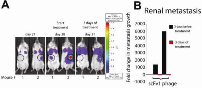
Figure 3. Examples of extrapulmonary lesion regression under treatment with scFv 1.
(A) F-luc tagged, in vivo selected and highly metastatic MDA-MB-435-met cells were injected i.v. to induce multiorgan metastasis. Metastatic progression was monitored by non-invasive bioluminescence imaging (photons/second/cm2) over time. In these examples, the location of renal lesions is circled and shown 3 days before treatment, at the beginning of treatment, and after 3 daily doses of scFv1 phage. (B) Fold-change in renal lesion growth, calculated based on signal change during 3 days before and 3 days under treatment.

