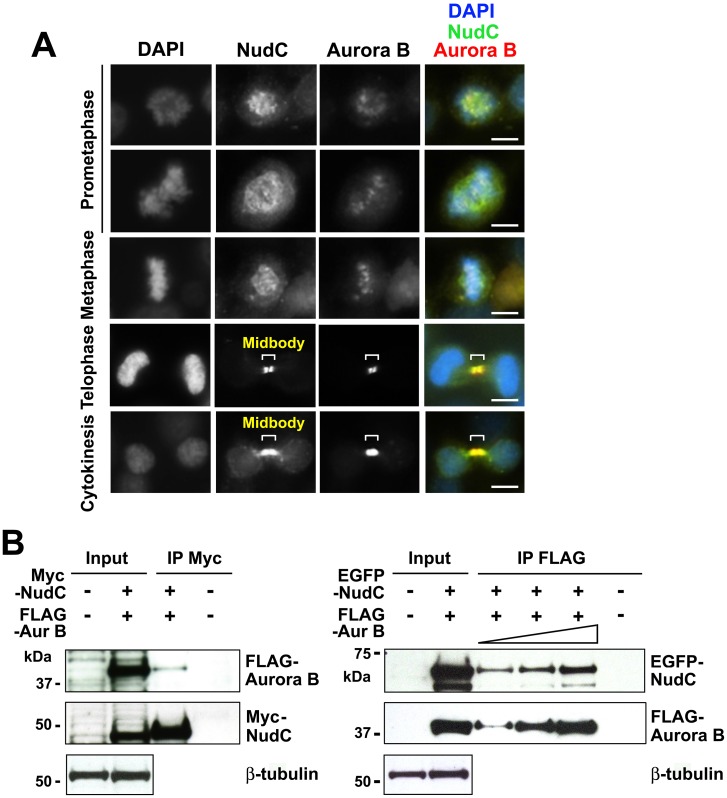
Fig 1. NudC co-localizes with Aurora B in mitosis.
(A) Unperturbed mitotic HeLa cells were stained for NudC (green), Aurora B (red) and counterstained with DAPI (blue). Bar, 10 μm. (B) HeLa cells were transfected with Myc-NudC and FLAG-Aurora B (left) or EGFP-NudC and FLAG-Aurora B (right) for 24 h. Cell lysates (1 mg in 250 μl) were immunoprecipitated with anti-Myc antibody and blotted for Aurora B followed by reblotting for NudC (left). A reciprocal immunoprecipitation was performed, in which cell lysates (500 μg in 250 μl, 1 mg in 250 μl or 2 mg in 500 μl) were immunoprecipitated with anti-FLAG antibody followed by blotting for NudC and reblotting for Aurora B (right). Immunoprecipitation with either anti-Myc or anti-FLAG antibody using non-transfected cell lysates was used as a negative control. β-tubulin was used as a loading control. Input, 20 μg total cell lysates. Data are representative of n = 5 independent experiments.

