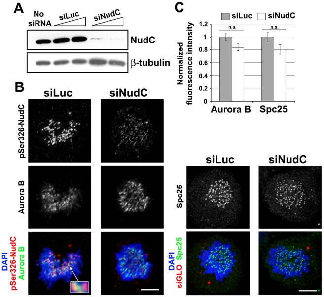
Fig 5. Aurora B localization at the kinetochore is not affected in NudC-deficient cells.
(A) HeLa cells were transfected with siLuc or siNudC oligos for 72 h. NudC knockdown was examined by western blotting for NudC. β-tubulin was used as a loading control. (B) Prometaphase cells treated with siRNAs as in (A) were stained for pS326-NudC (red) and Aurora B (green) (enlarged in inset), or with Spc25 (green), and counterstained with DAPI for DNA (blue). In initial experiments, siGLO was co-transfected as an indicator for siRNA oligo uptake. (C) For quantification, cells treated as in (B) were also co-stained with the CREST autoserum to mark the kinetochores. For Aurora B or Spc25 staining, maximum-intensity projections of deconvolved images were measured using AutoDeblur/AutoVisualize software, and their fluorescence intensities (average ± s.d.) relative to that of CREST staining at the kinetochore were quantified, using 10 randomly chosen kinetochores from at least 10 siLuc or siNudC prometaphase cells. n.s., not significant.

