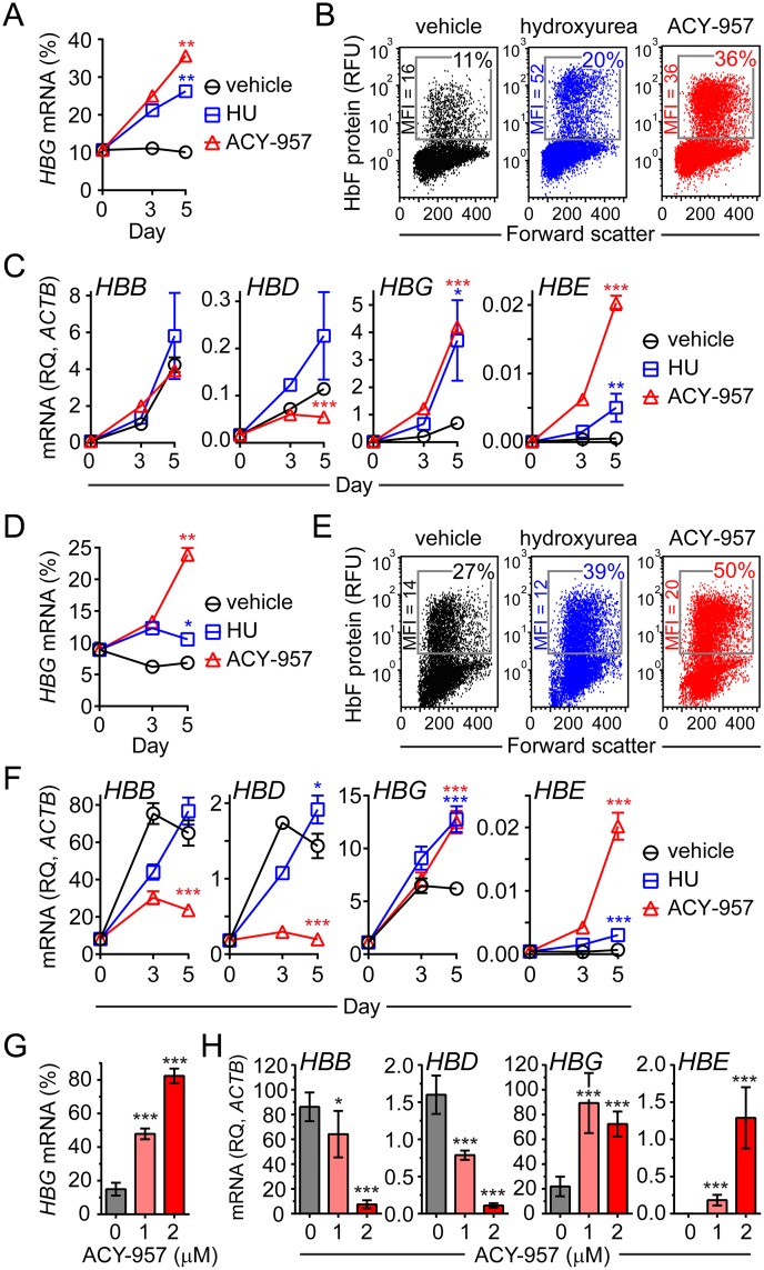
Fig 2. ACY-957 induces HBG mRNA and HbF protein in primary cells from healthy donors.
(A) Time-dependent increase in the percent HBG mRNA in CS1 cells. BM cells were cultured in CS1 expansion media, then shifted to CS1 differentiation media for 5 days with vehicle (dimethyl sulfoxide), 30 μM hydroxyurea (HU), or 1 μM ACY-957 (mean ± SD, n = 2 QPCR and n = 2 cell culture replicates). Data is representative of experiments using cells from three independent donors. (B) HbF protein induction in ACY-957 treated cells. Cells from day 5 of differentiation in ‘A’ were stained with an anti-HbF antibody and detected by flow cytometry. (C) Effect of ACY-957 on each β-like globin transcript. Samples from ‘A’ with each β-like globin transcript plotted relative to β-actin (ACTB). (D) Time-dependent increase in the percent HBG mRNA in CS2 cells. BM cells were cultured in CS2 expansion media, then shifted to CS2 differentiation media for 5 days with vehicle, 30 μM HU, or 1 μM ACY-957 (mean ± SD, n = 2 QPCR and n = 2 cell culture replicates). Data is representative of experiments using cells from two independent donors. (E) HbF protein induction in ACY-957 treated cells. Cells from day 5 of differentiation in ‘D’ were stained with an anti-HbF antibody and detected by flow cytometry. (F) Samples from ‘D’ with each β-like globin transcript plotted relative to ACTB. (G) Dose-dependent increase in percent HBG mRNA in BFU-E colonies derived from human bone marrow mononuclear cells cultured with ACY-957 (mean ± SD, n = 3 cell culture replicates). Data is representative of experiments using cells from two independent donors. (H) Samples from ‘G’ with each β-like globin transcript plotted relative to ACTB. In panels ‘A’, ‘C’, ‘D’, and ‘F’, P-values were calculated on day 5 only using a two-tailed t test. In panel ‘G’ and ‘H’ P-values were calculated using a two-tailed t test. For all panels *P<0.05, **P<0.005, and ***P<0.0005 compared to vehicle treatment. HBG mRNA (%) = [HBG/(HBB+HBD+HBG+HBE)]*100. MFI, mean fluorescent intensity. RFU, relative fluorescence units. RQ, relative quantity.

