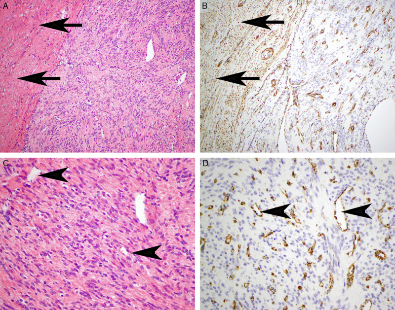
FIGURE 1.

Serial hematoxylin and eosin–stained (A, C) and FH IHC-stained (B, D) sections. The non-neoplastic uterine smooth muscle (arrows) and endothelial cells within the main tumor mass (arrowheads) demonstrate positive staining for FH. This staining, which is distinctly mitochondrial (granular and cytoplasmic), serves as an internal positive control and contrasts with the leiomyoma, which is completely negative. The positive staining of the endothelial cells also serves to highlight the hemangiopericytomatous and slit-like vascular pattern.
