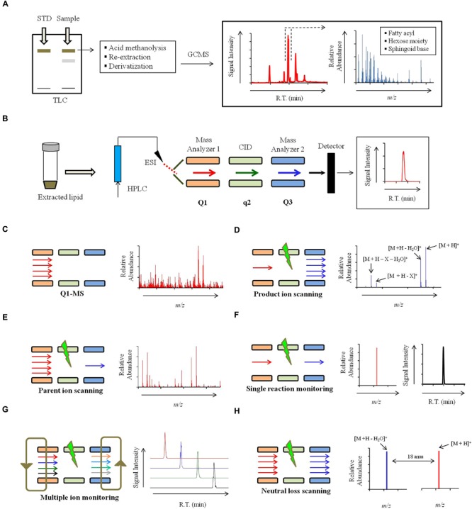
FIGURE 4.

Strategy for mass spectrometry based identification of sphinglipids. (A) Alternate strategies to confirm the sphingolipid structure. Qualitative analysis of sphingolipids can be performed using commercially available standards by High performance thin-layer chromatography (HP-TLC) or TLC. Sphingolipid components like sphingoid backbone, sugar and fatty can be analyzed using Gas chromatography mass spectrometry (GCMS). For sphingoid base and sugar analysis, samples are prepared using following steps: acid methanolysis (1 N HCl in methanol), N-acetylation (pyridine and acetic anhydride) and per-O-trimethylsilylation [TriSil reagent; 1-(Trimethylsilyl)imidazole – Pyridine mixture]. Sphingoid bases and sugars are analyzed as N-acetyl-per-O-trimethylsilyl derivatives and monosaccharide methyl glycosides, respectively. For fatty acyl analysis, samples are prepared using acid methanolysis, re-extraction in n-hexane and per-O-trimethylsilylated (TriSil) to form fatty acid methyl esters (FAMEs). (B) A scheme of HPLC-ESI-MS/MS. Lipids are resolved by HPLC and thereafter analyzed by tandem mass spectrometry (MS/MS). ‘Q1’ and ‘Q3’ quadruples are mass analyzers and the quadrupole ‘q’ operates in radio frequency leading to collision-induced dissociation (CID). Electrospray ionization (ESI) or Atmospheric pressure chemical ionization (APCI, not shown) is used as ionization methods. (C) Scheme for Q1-MS or full scan. The molecular ions or m/z ratio are recorded in Q1. (D) Scheme for product ion scanning. Only a select ion with specific m/z is recorded in Q1 and undergoes CID. Resultant ions are recorded in Q3. ‘M’ represents the molecular ion; ‘X’ represents a fragment loss. (E) Scheme for parent ion scanning. A full scan of ions recorded in Q1 and following CID, only a select fragment ion is recorded in Q3. (F) Scheme for single-reaction monitoring (SRM). A preselected ion is recorded in Q1, a select parent ion undergoes CID and a select fragment ion is recorded in Q3. (G) Scheme for multiple-reaction monitoring (MRM). Multiple SRM’s can be set up simultaneously depending upon the scanning efficiency of the instrument. (H) Scheme for neutral loss (NL) scanning. Ions are recorded in both Q1 and Q3 with a set mass difference between the two.
