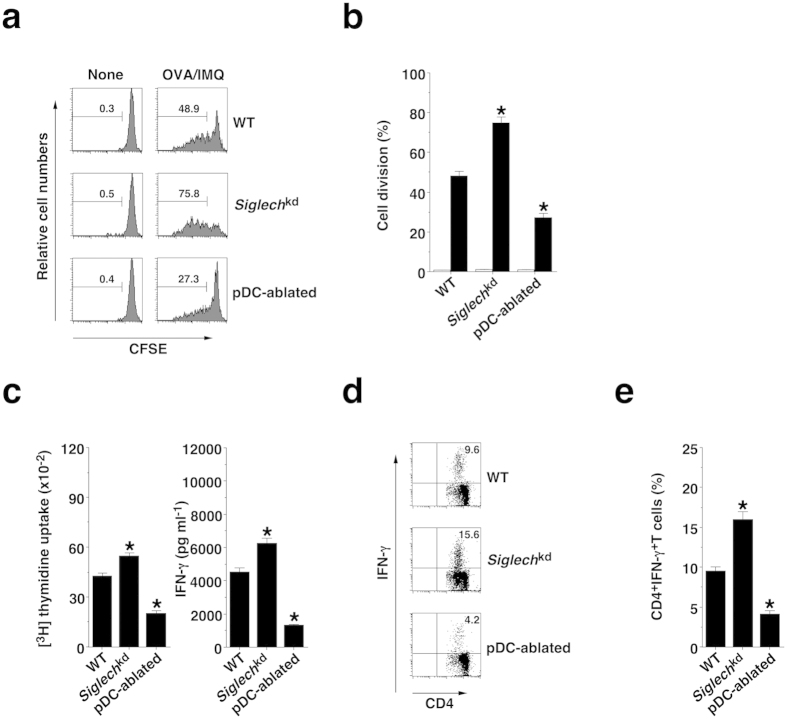
Figure 4. pDCs promote TLR7-mediated Ag-specific CD4+ T-cell responses in vivo.
(a,b) CFSE-labeled CD45.1+OT-II CD4+ T cells were transferred into the C57BL/6-background WT mice (n = 6), Siglechkd mice (n = 6), and pDC-ablated mice (n = 6), and then the mice were immunized with IMQ plus OVA protein. Ag-specific division of CD45.1+OT-II CD4+ T cells was analyzed 3 days after the immunization by flow cytometry. (a) Data are presented as a histogram, and numbers represent the proportion of the dividing cells among gated CD45.1+OT-II CD4+ T cells in each histogram. (b) Data are the mean percentage of positive cells ± s.d. from six individual samples in a single experiment. (c–e) The C57BL/6-background WT mice (n = 6), Siglechkd mice (n = 6), and pDC-ablated mice (n = 6) were immunized with OVA protein plus IMQ. At 14 days after the immunization, Spl CD4+ T cells were cultured with WT CD11c+ DCs in the presence or absence of OVA protein for the measurement of proliferative responses by [3H]thymidine incorporation ((c) left panel) and production of IFN-γ ((c) right panel) by ELISA. (d,e) Intracellular production of IFN-γ in the cultured CD4+ T cells was analyzed by flow cytometry. (d) Data are presented as a dot plot, and numbers represent the proportion of IFN-γ+ cells among gated CD4+ T cells in each quadrant. (e) Data are the mean percentage of positive cells ± s.d. from six individual samples in a single experiment. *P < 0.01 compared with WT mice. All data are representative of at least three independent experiments.

