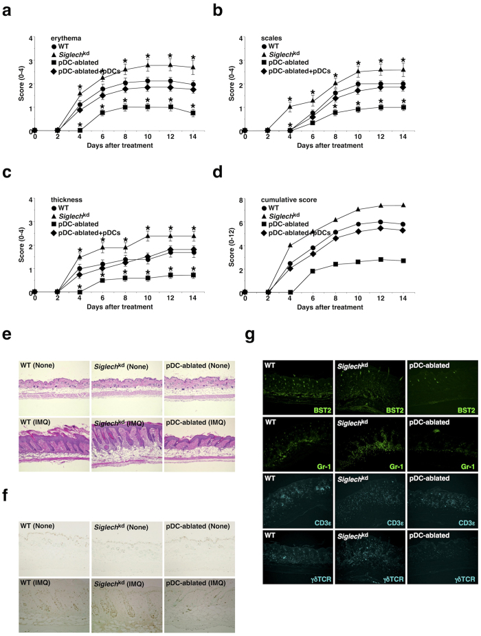
Figure 6. pDCs aggravate IMQ-induced psoriasiform dermatitis.
The C57BL/6-background WT mice (n = 10), Siglechkd mice (n = 10), and pDC-ablated mice (n = 10) that had been adoptively transferred with or without pDCs were treated topically with IMQ cream on the shaved back every other day for 14 days. (a–d) The disease severity of each mouse was scored daily, and erythema (a), scaling (b), thickness (c) and cumulative score (d) of the back skin at the indicated times were plotted. Data are the mean ± s.d. from ten individual samples in a single experiment. *P < 0.01 compared with WT mice. (e) H&E staining of the paraffin-embedded sections (magnification; 10×) obtained from the back skin of untreated mice (upper panel) and mice 6 days after treatment with IMQ cream (lower panel). (f) Immunohistochemical detection of proliferating cell nuclear Ag (PCNA) was performed on paraffin-embedded sections (magnification; 10×) obtained from the back skin of untreated mice (upper panel) and mice 6 days after treatment with IMQ cream (lower panel). (g) Immunofluorescent microscopic analysis was performed on frozen horizontal sections (magnification; 10×) obtained from the back skin of mice 6 days after treatment with IMQ cream. Sections were stained for BST2 (green), Gr-1 (green), CD3ε (cyan) or γδTCR (cyan). All data are representative of at least three independent experiments.

