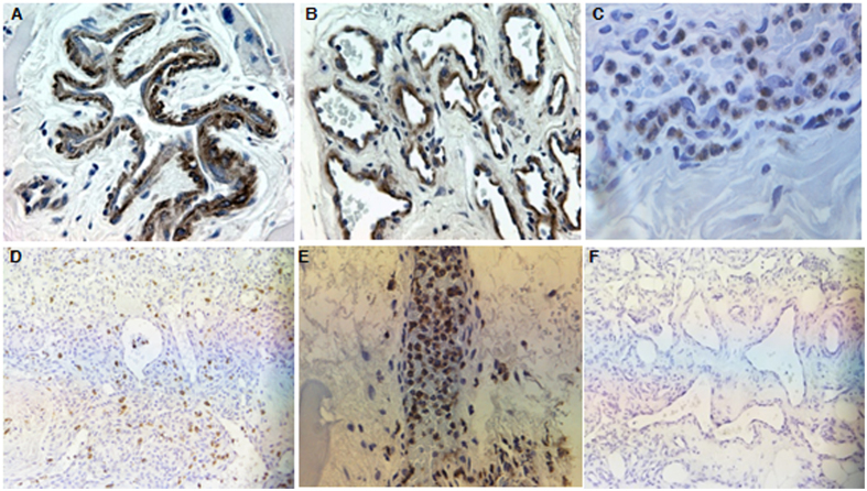
Figure 6. Immunohistochemistry analysis of MMP-9, NGAL and MMP-2 in AVMs.
(A–C), MMP-9 was predominantly located in perivascular cells, endothelial cells and neutrophils. (D,E), abundant NGAL positive neutrophils were widely scattered in AVM tissues. (F), MMP-2 staining was negative in all AVMs and normal tissues. Original magnification ×400 (A–C) and ×200 (D–F).

