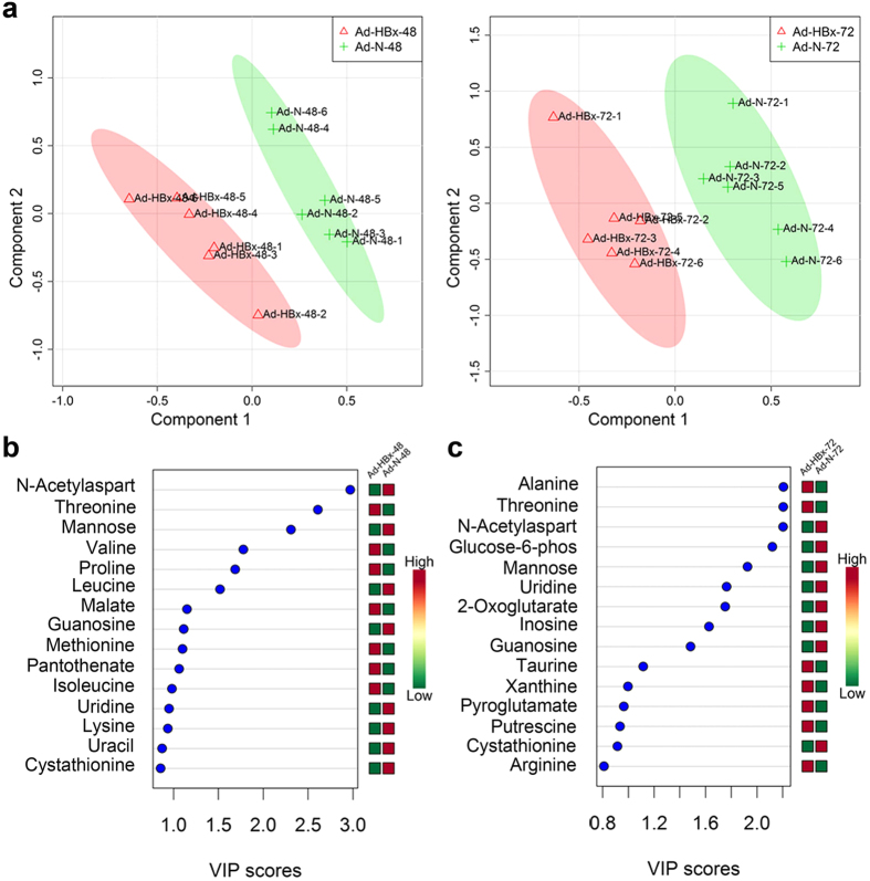
Figure 2. HBx-induced abnormalities of nucleic acids.
(a) Scores plots of Ad-HBx-48 (red triangles) and Ad-N-48 (green crosses), and Ad-HBx-72 (red triangles) and Ad-N-72 (green crosses). (b) VIP scores of Ad-HBx-48 and Ad-N-48. (c) VIP scores of Ad-HBx-72 and Ad-N-72. Red or green on the right of (b) and (c) indicates the low or high concentration of metabolites by comparing the concentration of each metabolite in Ad-HBx-48 and Ad-N-48 or Ad-HBx-72 and Ad-N-72, respectively.

