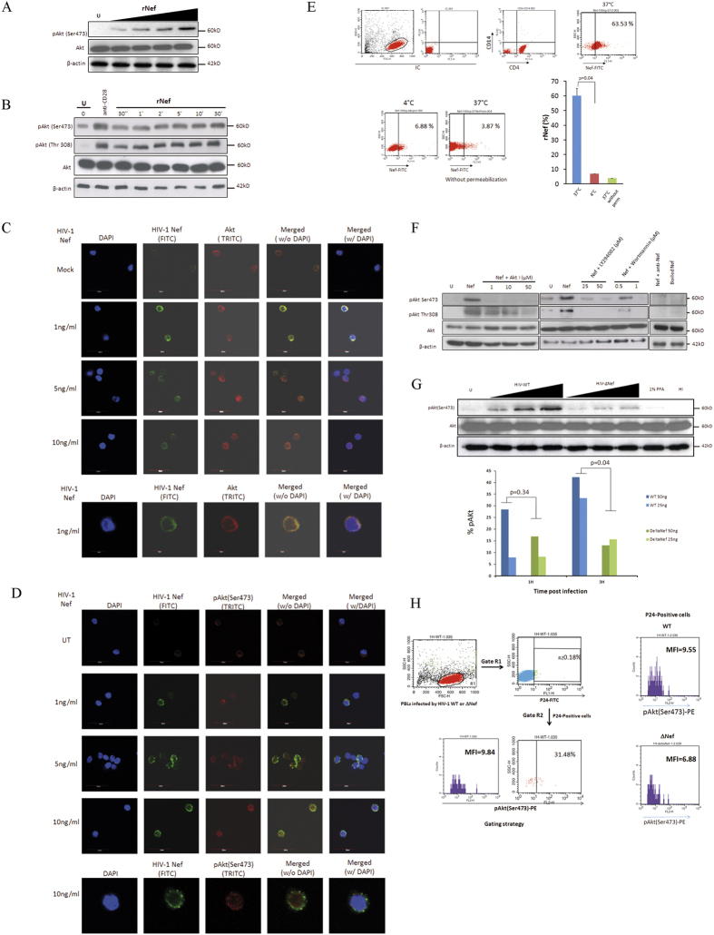
Figure 1. HIV-1 Nef is internalized by CD4 + T cells and activates Akt in PBLs which is mediated via PI3K in a dose and time dependent manners.
(A), Dose-dependent (n = 3) and (B), Time-dependent activation of Akt (pAkt(Ser473)) in PBLs treated with rNef (n = 3). (B) Five million PBLs were either left untreated or treated with rNef (100 ng/ml) for various period of time (30 seconds to 30 minutes). As a positive control, PBLs were treated with anti-CD28 antibody. Expression of pAkt (Ser473, Thr308), total Akt and β-actin were detected by standard western blotting method as described in materials and methods (n = 3). (C,D) A series of confocal images showing internalization and colocalization of HIV-1 Nef and Akt (C) at serum concentration of Nef (1 to 10 ng/ml) and a dose response of rNef treatment on Akt activation (D) in PBLs isolated from healthy donors. (E), Internalization of rNef by CD4+ T cells determined by flow cytometry. Five million CD4+ T cells were treated with rNef for 30 min at 37 °C and 4 °C with and without permeabilization. Expression of rNef was determined by confocal microscopy (n = 3).(F), Activation of Akt in PBLs treated with rNef is mediated via PI3K. Western blot detection of activated pAkt (Ser473, Thr308) in the lysates derived from 5 × 10⁶ PBLs treated with 100 ng/ml of Nef with or without Akt (Akt inhibitor VIII) and PI3K inhibitors (LY294002 and Wortmannin) (n = 3). (G), Akt activation in PBLs by wild-type HIV-1, but not by isogenic HIV-1∆Nef. Five million PBLs were infected with various doses (of p24) of either wild type HIV-1 or ∆Nef virus infectious clones. Post infection lysates were prepared and expression of pAkt(Ser473), was determined by western blotting (upper panel) and flow cytometry (n = 2) (lower panel). (H), Left panel: Gating strategy for flow cytometry analysis. In this sample gating cells were first gated for PBLs (Gate R1). R1 gate is further analysed for p24 positive PBLs (Gate R2). Gate R2 is analyzed for the expression of pAkt. Right panel is the representative data of pAKt positive cells in p24 positive population obtained by infection of PBLs with WT and Delta Nef virus.

