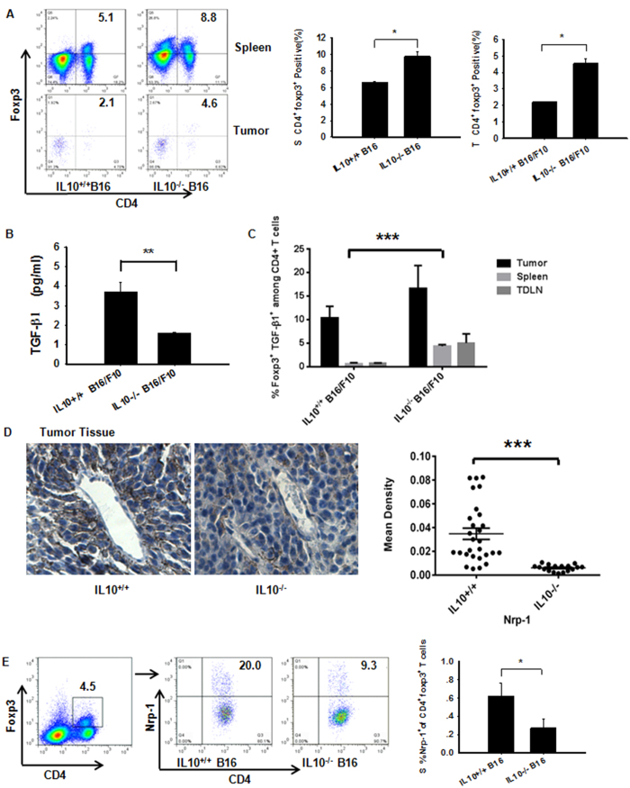
Figure 4. Over-compensation of IL-10 abrogation leads to impairment of treg-Derived Nrp-1 but not TGF-β1 production in B16/F10 tumors.
(A) Implantation of IL-10-deficient mice with B16/F10 cells resulted in enhanced CD4+Foxp3+ Tregs in the spleen and tumor on day +15, as assessed by flow cytometry. (B) Measurement of TGF-β1 in NP-40-processed B16/F10 tumor tissues (described in the Methods section) obtained from WT and IL-10-deficient tumor-bearing mice. The TGF-β1 cytokine was measured using Luminex (Millipore Merck). (C) Among CD4+ T cells from IL10−/− tumor-bearing mice, splenic TGF-β1+Foxp3+ cells were increased compared with WTB16/F10 mice; however, differences in the tumor tissues and TDLNs were not significant. (D) IL10 deficiency down-regulated Nrp-1 protein expression in tumor tissues. Representative photomicrographs of B16/F10 tumors harvested on day 15. Left panel, representative immunohistochemistry images obtained using a microscope with a 20X objective. Right panel, statistical analysis of the mean IOD for tumor cell proliferation using Image-Pro Plus software. Nonlinear regression was performed using GraphPad Prism (San Diego, CA, USA). (E) Spleen Nrp-1-expressing CD4+Foxp3+ T cells from IL10−/− tumor-bearing mice were decreased compared with WT B16/F10 mice. Panels (A,B,D,E) Evaluated using Student’s t-test to determine statistical significance with *P < 0.05, **P < 0.01, ***P < 0.001between experimental groups. The data are expressed as the mean values ± SEM from n = 3–4 mice. Panel (C) Evaluated using ANOVA, ***P < 0.001, ±SEM from n = 3–4 mice.

