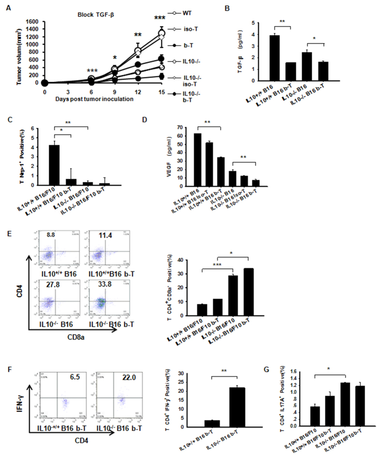
Figure 7. IL-10 but not TGF-β plays the primary role in Th1 and Th17 expansion in B16/F10 tumors and Peripheral Immune Organs.
(A) A neutralizing anti-TGF-β antibody (described in the Methods section) was used to treat WT or IL10−/− B16/F10 tumor-bearing mice. The tumor mass volume was monitored every three days after implantation. (B) TGF-β1 from NP-40-processed B16/F10 tumor tissues was measured using Luminex. Dual inhibition of IL-10 and TGF-β resulted in significantly lower levels of tumor-secreted VEGF protein in B16/F10 tumor-bearing mice. (C) Tumor-derived WT B16/F10-derived Nrp-1 levels were elevated compared with IL10−/− B16/F10 mice treated with anti-TGF-β or IL10−/− B16/F10 mice by flow cytometry. (D) VEGF from NP-40-processed B16/F10 tumor tissues measured using Luminex. (E) The absence of IL-10 or blocked TGF-β antibody had a more pronounced effect on CD4+ CD8a− T cells in B16/F10 tumors. (F) Anti-TGF-β in IL10−/− B16/F10 mice allowed for the expansion of tumor IFN-γ-expressing CD4+ (Th1) populations measured by flow cytometry. (G) Anti-TGF-β did not allow the expansion of tumor CD4+ IL17A+ (Th17) populations in IL10−/− B16/F10 mice measured by flow cytometry. The data shown are from one representative experiment of two independent experiments with same results and were evaluated using ANOVA for the determination of statistical significance with *P < 0.05, **P < 0.01, ***P < 0.001between experimental groups. The data are expressed as the mean values ± SEM from n = 4–6 mice.

