Fig. 3.

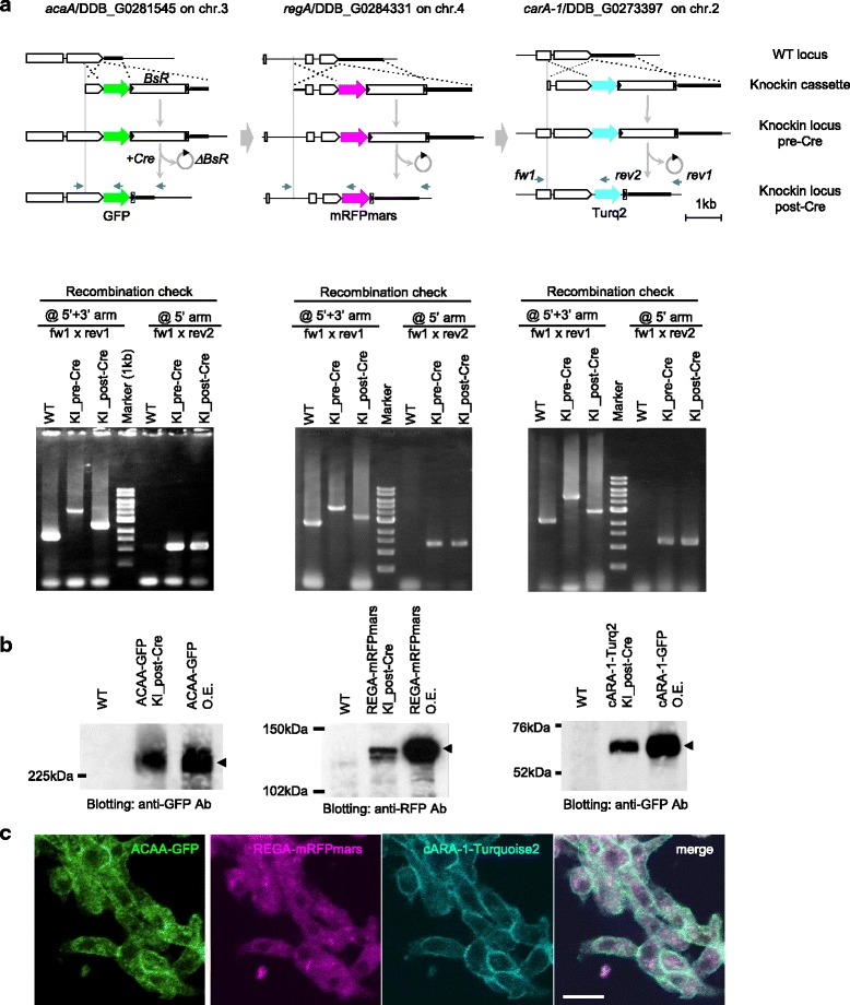
Triple-colour knockin for ACAA-GFP, REGA_mRFPmars and cARA-1-Turq2. a Genomic organization of wild-type (WT) and knockin locus (KI) for DDB_G0281545/acaA (left column), DDB_G0284331/regA (middle column) and DDB_G0273397/carA-1 (right column) tagged with green, red and cyan fluorescent proteins, respectively. Serial knockin in this order was performed, each followed by Cre-mediated BsR-recycling. Specific recombination was detected by PCR as in Fig. 2b. Primer fw1/rev1 detects a knockin event (WT to knockin_pre-Cre) and removal of BsR cassette (knockin_pre-Cre to post-Cre) as a 2.5 kb increase and 1.7 kb decrease of the amplified band, respectively. The fw1/rev2 set detects specific recombination at the 5′ recombination arm. b Protein expression of triple-colour knockin strain. Lysate from over-expressing cells (1/10 volume) with ACAA-GFP, REGA-mRFPmars and cARA-1-GFP were loaded as the positive control. c Live confocal images of the triple-colour knockin strain developed for 8 h. ACAA-GFP and cARA-1-Turq2 were localized at the cell surface. REGA-mRFPmars was detected in the cytoplasm excluded from the nucleus. Bar: 10 μm
