
Fig. 3.
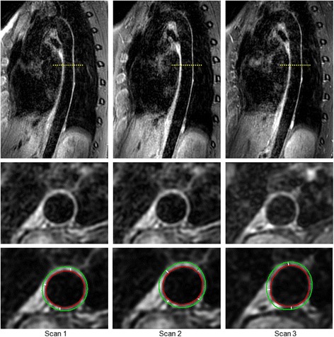
Example of 3D-T1-BB-VISTA acquisition. Examples of the original sagittal and reconstructed plain and traced transverse images of the descending thoracic aorta obtained with the 3D-T1-BB-VISTA sequence in a 32 year old female participant. Acquisitions 1 and scan 2 are identical non-contrast enhanced acquisitions at different time points. Acquisition 3 is a post-contrast acquisition. Aortic wall thicknesses for this slice were 1.52 mm, 1.53 mm and 1.51 mm respectively