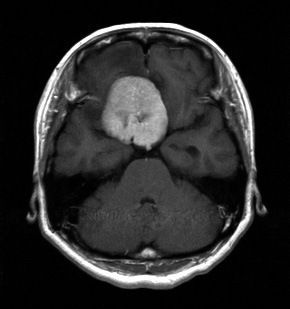
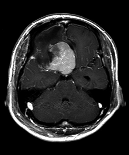
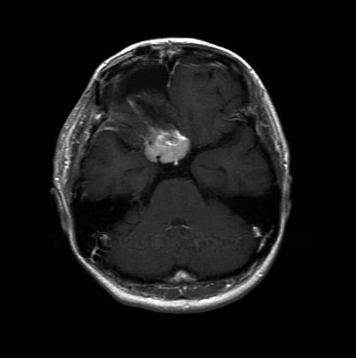
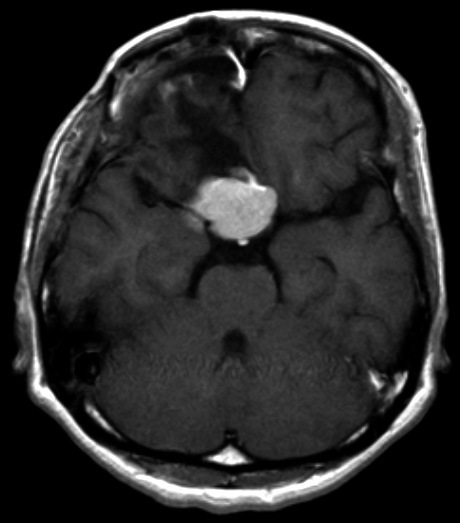
Fig. 2.
Gd-DTPA enhanced MRI, axial view
Fig. 2A.

MRI on admission. Tumor was located at the tuberculum sellae.
Fig. 2B.

MRI after first operation. Tumor was only partially resected.
Fig. 2C.

MRI after second operation. Tumor was diminished remarkably compared to preoperative size.
Fig. 2D.

MRI 25 months after second operation. Some enlargement of tumor was seen in comparison with size after second operation.
