Fig. 2.

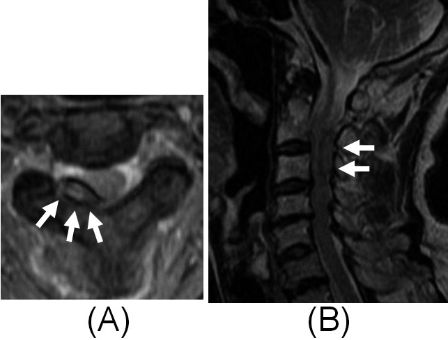
Magnetic resonance images of the cervical spine which were obtained on the third day after the onset. Axial (A) and sagittal (B) T2WIs show an extradural mass with hyperintensity that compressed the spinal cord (arrows).

Magnetic resonance images of the cervical spine which were obtained on the third day after the onset. Axial (A) and sagittal (B) T2WIs show an extradural mass with hyperintensity that compressed the spinal cord (arrows).