Figure 1.
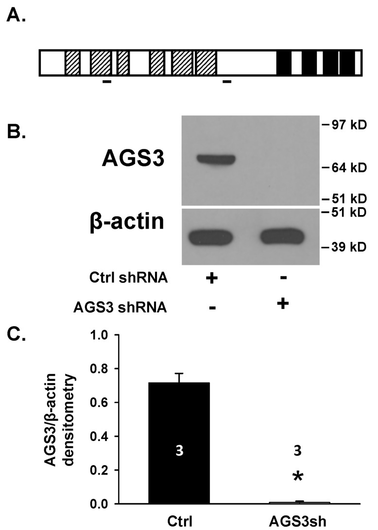
Genetic modification of NRK epithelial cells using lentiviral vectors to reduce endogenous levels of AGS3. (A) Schematic of AGS3 protein structure. Hatched bars = TPR motifs; solid black = G-protein regulatory (GPR) domains; solid lines = AGS3 shRNA target sites. (B) Protein lysates from NRK-52E cells transduced with lentiviral vectors expressing either control (NRK-Ctrl) or two distinct AGS3-specific shRNA (NRK-AGS3sh) were isolated for immunoblot analysis using a polyclonal AGS3 antibody. β-actin was shown as a loading control. Protein standards are shown on the right (in kD) for each blot image. (C) Graphical analysis of the AGS3 band intensity. Three different samples were used in each group, and were shown in the bars. * P < 0.001 significant difference between NRK cells expressing Ctrl versus AGS3-specific shRNA.

