
Figure 1.
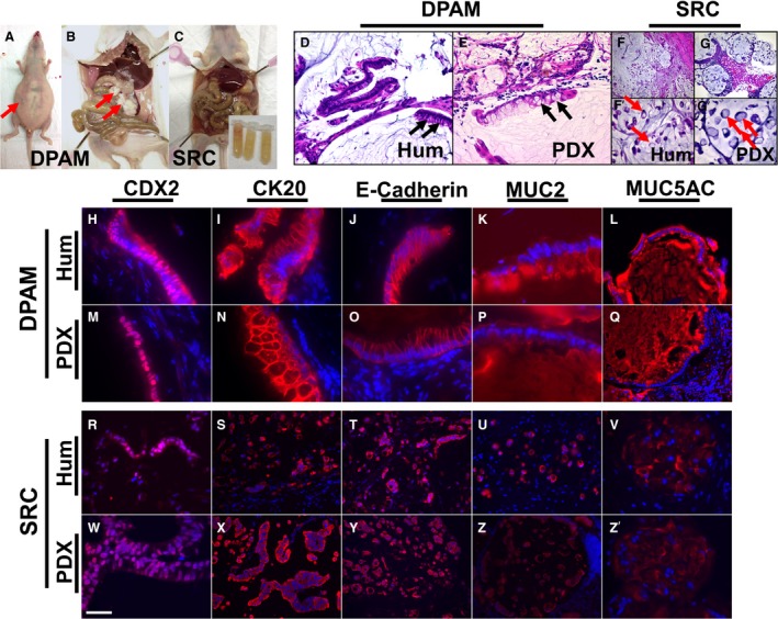
Gross morphological (A–C), histological (D–G’), and immunohistochemical (H–Z’) characteristics of PMP tumors in PDX models were similar to human PMP. Arrows in (A and B) point to a distended abdomen filled with ascites and tumor implants on serosal surface of visceral organs respectively. Arrows in (D and E) point to goblet cells. Arrows in (F’ and G’) point to signet ring cells. Inset in (C) (bottom right) shows tubes filled with ascites collected from the mouse. In (H–Z’), antigen‐antibody complexes are in red and nuclei are stained blue with DAPI. DPAM, disseminated peritoneal adenomucinosis; Hum, human; PDX, patient‐derived xenograft; SRC, signet ring cells. Scale bar: 60 μm in (F and G); 30 μm in (D, E, L, Q, S–U, X–Z); 15 μm in (F’, G’, R, W, V, and Z’); 10 μm in (H, I, J, K, M–P).