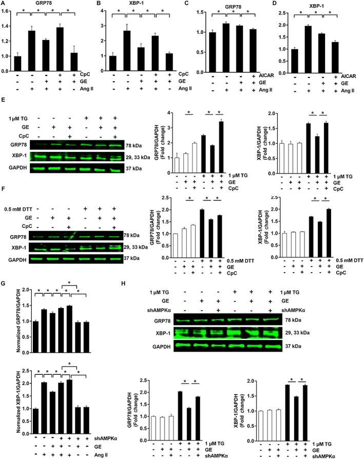
Figure 4.

The effects of geniposide (GE) on ER stress were blunted by CpC or shAMPKα in vitro. (A–B) The effects of geniposide (100 μM for 6 h) on ER stress were abolished by CpC in H9c2 cells (20 μM for 6 h) (n = 6). (C–D) The synergistic effects of geniposide and AICAR on ER stress in H9c2 cells (1 mmol · L−1 for 24 h) (n = 6). (E–F) ER stress induced by TG (1 μM for 6 h) or DTT (1 μM for 6 h) (n = 10). (G) ER stress induced by angiotensin (Ang II, 1 μM for 24 h) was abolished by shAMPKα in neonatal rat cardiomyocytes (n = 5). (H) ER stress induced by TG was abolished by shAMPKα in neonatal rat cardiomyocytes (n = 6). * P < 0.05.
