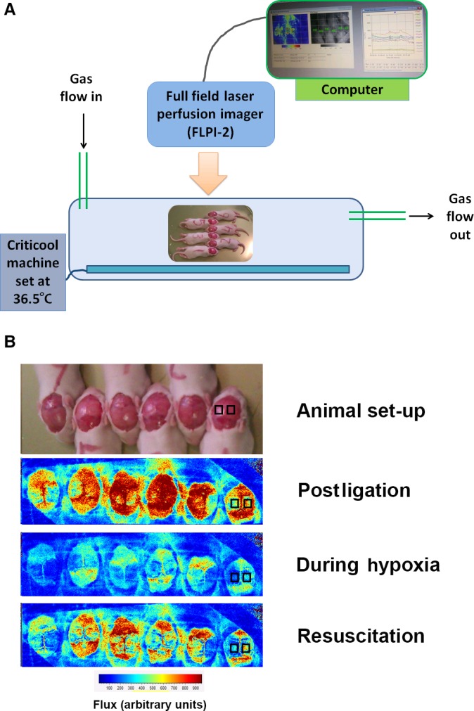
Figure 1.

Laser speckle imaging. Experimental set‐up (A) and representative images (B) using full‐field LSI to determine hemispheric blood flow (measured in arbitrary units of “flux”). The lower panel shows six animals with scalps exposed, within the hypoxia chamber. Animals faced in alternating directions to minimize breathing restrictions. Cerebral blood flux measurements (1) after ligation; (2) during hypoxia; and (3) after 10 min of resuscitation in 21% oxygen are shown. Representative bilateral regions of interest over the ligated (L) and unligated (R) hemispheres are shown in the animal on the right.
