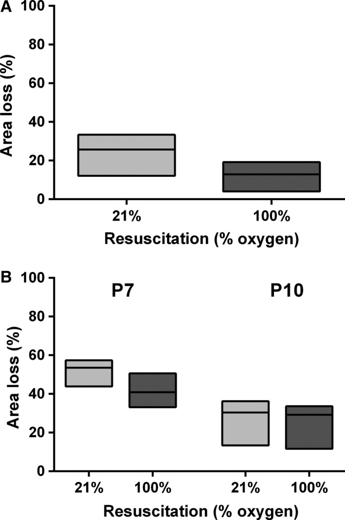
Figure 3.

Hemispheric area loss after resuscitation in 21% or 100% oxygen. (A) Area loss after control flux measurements. Box plot (Hodges–Lehmann median with 95% CI) of hemispheric area loss in P10 rats after unilateral HI under anesthesia, and sham CBF measurements, before resuscitation in either 21% oxygen (n = 11) or 100% oxygen (n = 13). (B) Area loss after hypoxia‐ischemia in P7 and P10 rats. Box plot (Hodges–Lehmann median with 95% CI) of hemispheric area loss in P7 and P10 rats after unilateral HI and resuscitation in either 21% oxygen or 100% oxygen.
