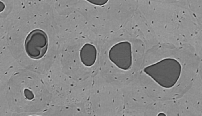
Figure 1.

Synchrotron micro‐CT scanning creates a cross‐sectional view of several osteons under digital zoom. Reversal lines were traced on each 10th slice, with all slices available for interpretation of changing morphology along the z‐axis. If reversal lines were slightly obscured in one image, their morphology could be easily determined by altering the viewing perspective up and down along the volume's depth (z‐dimension).
