
Figure 3.
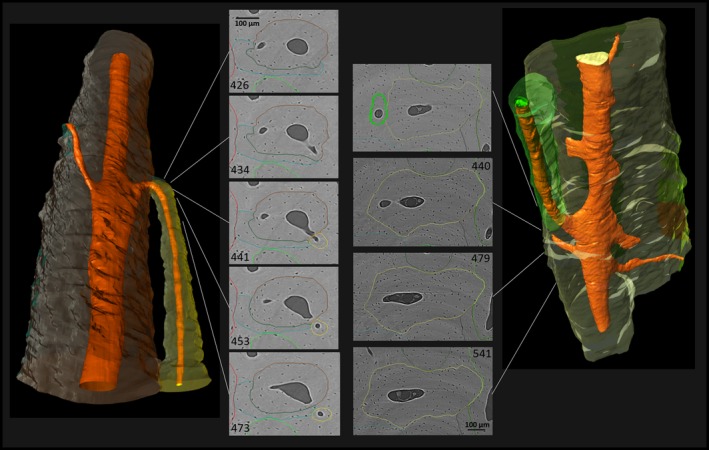
Lateral branching characterized by a newly formed canal that connects the Haversian canal of a previously existing Haversian system with adjacent tissue, where it extends through the formation of a new BMU. Note the consistency in cross‐sectional size and shape of the previously existing system, the almost perpendicular angle of the newly formed canal, especially in the left example, and the size difference between the ‘newer’ and the previously existing Haversian system. (For more information on image acquisition, see Fig. 1; numbers on 2D images indicate slice numbers; scale bar: 100 μm).