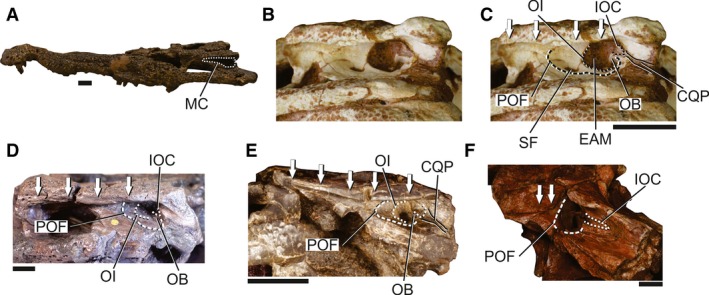
Figure 8.

Skull of neosuchians to illustrate the MC in these forms. (A) Overview of the skull of Amphicotylus lucasii (AMNH FARB 5782) in left lateral view to illustrate the general morphology of the MC in neosuchians. (B) Detail of the posterior portion of the skull and MC of Shamosuchus djadochtaensis (IGM 100/1195). (C) Detail of the posterior portion of the skull and MC of Shamosuchus djadochtaensis (IGM 100/1195), with selected structures of the MC highlighted. (D) Detail of the posterior portion of the skull and MC of Anterophthalmosuchus hooleyi (NHMUK R 3876), with selected structures of the MC highlighted. (E) Detail of the posterior portion of the skull and MC of Hylaeochampsa vectiana (NHMUK R 177), with selected structures of the MC highlighted. (F) Detail of the posterior portion of the skull and MC of Aegisuchus witmeri (ROM 54530), with selected structures of the MC highlighted. Arrows indicate the extension of the sulcus for the attachment of the upper earlid. CQP, cranioquadrate passage; EAM, external auditory meatus; IOC, incisure of the otic aperture of the cranioquadrate passage; MC, meatal chamber; OB, otic buttress; OI, otic incisure; POF; periotic fossa; SF, subtympanic foramen. Scale bars: 2 cm.
