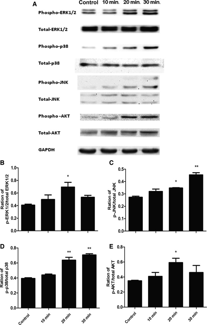
Figure 2.

Activation of ERK1/2, p38, JNK and AKT induced by IL‐1β in lung cancer cells (A549 cells). (A) Phosphorylated and total amounts of ERK1/2, p38 MAPK, JNK and AKT measured by Western blot in A549 cells; Ratio of p‐ERK1/2/total ERK1/2 (B), p‐p38/total p38 (C), p‐JNK/total JNK (D) and p‐AKT/total AKT (E) in A549 cells. Starved cells were challenged without (control) or with IL‐1β at 1 ng/ml for 10, 20, 30 min. Data were presented as mean ± S.E. and each group has at least six measurements (*P < 0.05, **P < 0.01).
