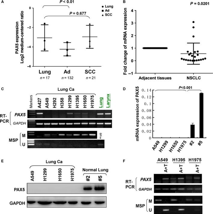
Figure 1.

The expression and methylation status of PAX5 in NSCLC. (A) PAX5 expression (median of expression intensity) in NSCLC from Oncomine database (https://www.oncomine.org/). Ad: lung adenocarcinoma; SCC: squamous‐cell lung carcinoma. (B) PAX5 expression in primary NSCLC tissues and paired adjacent non‐tumour tissues was evaluated using real‐time PCR (n = 23). PAX5 expression level was normalized with the mRNA level. (C) PAX5 expression by semi‐quantitative PCR and methylation status of the PAX5 promoter by MSP in a panel of human NSCLC cell lines and the PAX5 expression of normal lung and larynx tissue. β‐actin was used as a control. +Ve, positive control; M: methylated; U: unmethylated. (D) PAX5 mRNA expression in four NSCLC cell lines and two normal lung tissue samples by qRT‐PCR. β‐actin was used as a control. (E) PAX5 protein expression in four NSCLC cell lines and two normal lung tissue samples by western blot. GAPDH was used as a control. (F) Pharmacological demethylation restores expression of PAX5. A549, H1395 and H1975 cells were treated with Aza combined with TSA (A+T). PAX5 expression by semi‐quantitative PCR and methylation status of the PAX5 promoter by MSP after A+T treatment. GAPDH was used as a control.
