Figure 4.

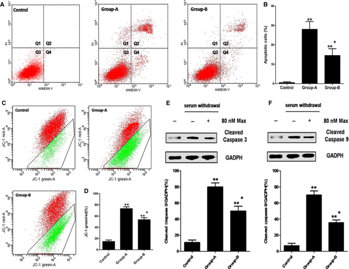
The anti‐apoptotic effects of maxadilan on hADSCs after serum withdrawal. (A) Annexin V and PI assays performed to detect the apoptotic of hADSCs in control, group‐A and group‐B by flow cytometry. Q1 area represents cell necrosis; Q2 is late‐apoptotic cells; Q3 is viable cells; Q4 is early‐apoptotic cells. (B) Quantification of the Annexin V and PI assays. (C) Mitochondrial membrane potential assays performed to assess the apoptotic of hADSCs in these groups. (D) Quantification of mitochondrial membrane potential assays. (E) Assessment of the expression of Cleaved Caspase 3 of hADSCs in these groups using Western blot assays. (F) Detection of the expression of Cleaved Caspase 9 of hADSCs in these groups using Western blot assays. Differences with **P < 0.01 (group‐A or group‐B versus control), *P < 0.05 (group‐B versus group‐A) were considered significant.
