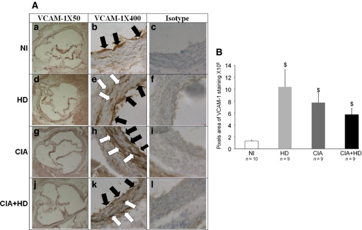
Figure 4.

VCAM‐1 staining in aortic sinus from mice fed an HD or standard chow diet. (A) Representative histological sections of the aortic sinus stained with blue‐haematoxylin for VCAM‐1 in NI mice fed a standard chow diet or an HD and in immunized mice fed a standard diet (CIA) or an HD (CIA+HD). Magnification is 50 × (a, d, g, j) or 400 × (b, e, h, k). Staining for isotype control of VCAM‐1 antibody (IgG2a, κ antibody) is shown in c, f, i and l (×400 magnification). Black arrows show VCAM‐1 endothelial staining; white arrows show VCAM‐1 staining in the aortic sinus wall. (B) Quantification of VCAM‐1–positive staining by use of Image J Fiji. $ P < 0.05 versus NI.
