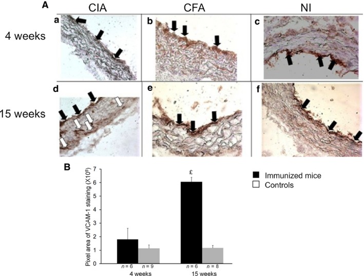
Figure 5.

Early (4 weeks) and late (15 weeks) VCAM‐1 staining in aortic sinus of mice. Aortic sinuses from CIA mice were removed at 4 and 15 weeks after immunization. Controls were immunized with only CFA or were NI mice. (A) Representative microphotographs of VCAM‐1 staining in aortic sinus from cCII/CFA‐immunized (CIA: a, d), CFA‐immunized (CFA: b, e) and NI(c, f) mice. Black arrows show endothelial staining for VCAM‐1; white arrows show VCAM‐1 staining in the aortic sinus wall. (B) Quantification of VCAM‐1–staining at 4 and 15 weeks in CIA versus control mice (NI and CFA mice pooled) by use of Image J Fiji. £ P < 0.05 versus CT.
