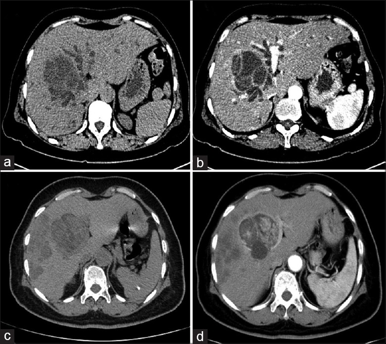
Figure 1.

(a) and (b) Computed tomography (CT) shows intrahepatic biliary cystadenoma in liver segment IV with intrahepatic bile duct dilation due to tumor compression; thin internal septa were observed in the tumor; (c) and (d) Intrahepatic biliary cystadenocarcinoma located in the left hepatic lobe with multiple metastases in the right lobe; internal septa and mural nodules showed mild contrast enhancement on venous phase CT.
