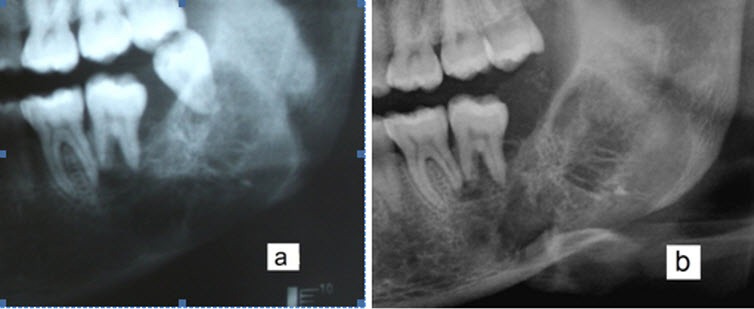
Figure 1.

Cropped panoramic radiograph showed a mixed radiolucent/radiopaque lesion with a relatively well-defined border between the left mandibular first molar and ramus. Before extraction (a). Six months after the extraction of third molar (b). Tooth displacement and root resorption were also seen on the panoramic radiograph.
