Figure 2.

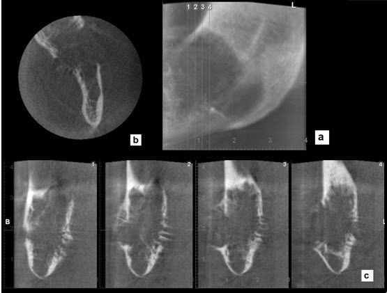
Panoramic (a) axial (b) and cross sections (c) showed perforation of the cortical plate and displayed septa at the periphery of the lesion.

Panoramic (a) axial (b) and cross sections (c) showed perforation of the cortical plate and displayed septa at the periphery of the lesion.