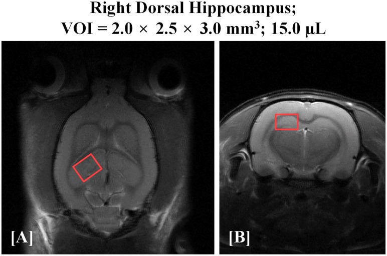
Fig 1. T2-weighted, FSE MR images [(A) coronal and (B) axial] of the rat brain with the volume of interest (VOI) centered in the right dorsal hippocampal region.
The red color illustrations on T2-weighted images indicate the size of the rectangular volume of interest as 15.0 μL. FSE: fast spin echo.

