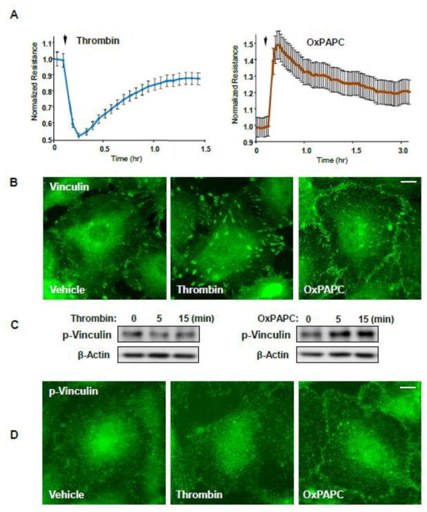
Figure 1. Effects of thrombin and OxPAPC on endothelial permeability and vinculin distribution.
A - HPAEC plated on microelectrodes were treated with thrombin (0.2 U/ml) or OxPAPC (15 μg/ml) followed by TER measurements. The TER curves represent pooled data from three independent experiments. B - Cells grown on glass coverslips were stimulated with thrombin or OxPAPC for 15 or 30 min, respectively. Vinculin localization was analyzed by immunofluorescence staining with corresponding antibody. Results are representative of three independent experiments. C - Western blot analysis of phospho-Y822 vinculin levels in HPAEC stimulated with thrombin (0.2 U/ml) or OxPAPC (15 μg/ml) at the indicated time points. D - Cells grown on glass coverslips were stimulated with thrombin or OxPAPC for 5 or 15 min, respectively. Phospho-Y822 vinculin localization was analyzed by immunofluorescence staining with corresponding antibody. Bar - 5 μm. Results are representative of three independent experiments.

