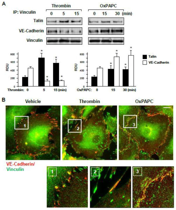
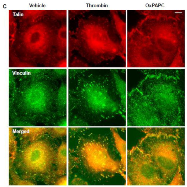
Figure 2. Effects of thrombin and OxPAPC on focal adhesion and adherens junction remodeling.
A – EC were stimulated with thrombin (0.2 U/ml) or OxPAPC (15 μg/ml), followed by vinculin immunoprecipitation under non-denaturing conditions. Presence of talin and VE-cadherin in immune complexes was tested by Western blot. Bar graphs depict quantitative analysis of Western blot data; n=3; *P<0.05 vs. vehicle. B and C – Thrombin- or OxPAPC-induced vinculin redistribution and its colocalization with VE-cadherin (B) and talin (C) was evaluated by immunofluorescence staining of formaldehyde-fixed EC for vinculin (green) and VE-cadherin or talin (red). Bar - 5 μm. Higher magnification insets show details of vinculin and VE-cadherin co-localization (yellow). Results are representative of three to five independent experiments.


