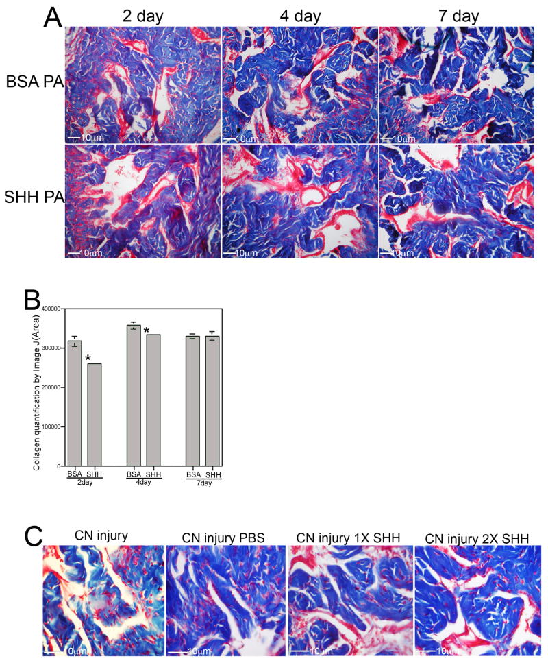
Figure 7.
Trichrome stain of penis tissue of rats that under went CN resection and SHH or BSA treatment by peptide amphiphile delivery to the penis (250X magnification). (B) Quantification of collagen by Image J analysis showed decreased collagen in CN injured penis with SHH treatment at 2 days (19% decrease, p=0.002) and 4 days (7% decrease, p=0.039) after injury/treatment. By 7 days SHH protein had been expelled from the PA and there was no difference in collagen abundance (p=0.485). (C) SHH protein delivery by Affi-Gel bead injection to the penis showed comparable results to peptide amphiphile delivery with unchanged corpora cavernosal morphology 4 days after CN resection in the presence of exogenous SHH protein.

