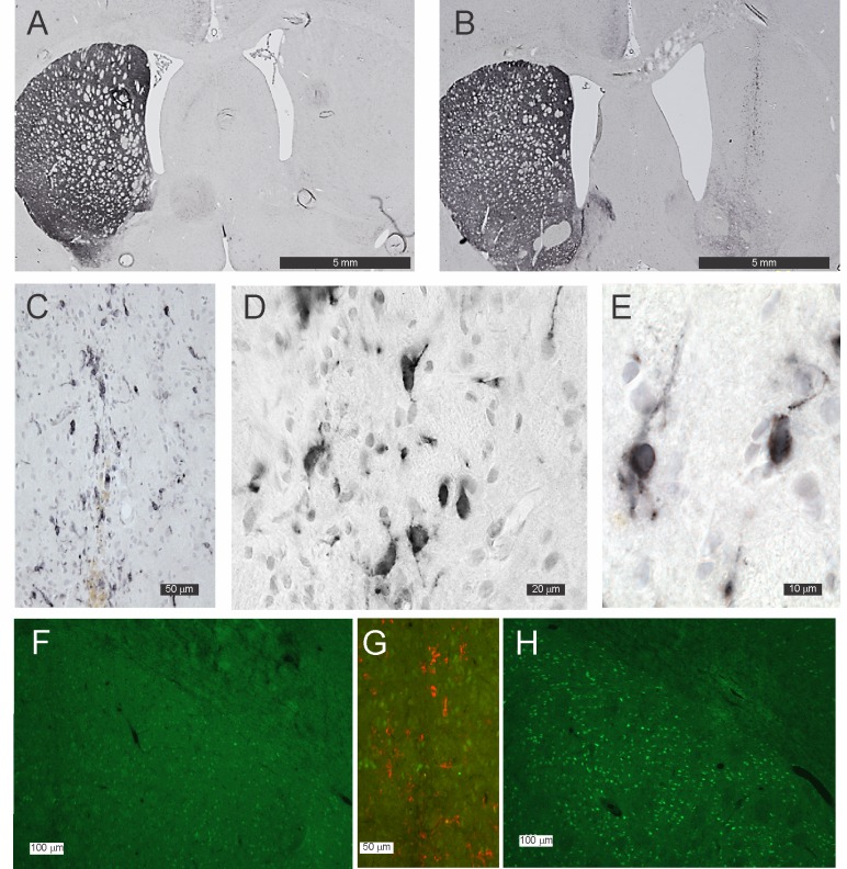
Fig 2. Photomicrographs of DAT-immunoreactivity in the striatum of grafted animals.
(A) Null graft located in the striatum of a rat in whom 6-OHDA had been used to denervate the striatum. The graft tract is faintly discernible but there are no DAT-immunoreactive cell bodies, terminals or fibres. (B, C, D and E) show a DAT graft located in the striatum of a rat in whom 6-OHDA had been used to denervate the striatum; note the numerous DAT-immunoreactive cell bodies within the graft (B and D). (D and E) show DAT-immunoreactivity is present in the cytoplasm and along the numerous processes emanating from the cells. (F, G and H) show Fos- B immuno-reactivity (green) in a DAT grafted striatum (F) and a Null grafted striatum (H). (G) shows both Fos_B and DAT immuno-reactivity (red) merged in the one image in the region of the DAT graft shown by the white rectangle in (F).

